How many small pictures become one big picture
Stitching as a technical term in image processing technology, it means that individual images are stitched together to form a panorama. In radiology, stitching has become an indispensable tool for creating special images and optimising images - both in human and veterinary medicine.
In precise terms, stitching is the process of seamlessly combining several images or image sections into a single, coherent image. This process is typically computerised and can be performed both manually and automatically. For reasons of dose optimisation, the overlapping area should be kept as small as possible.
Stitching is based on recordings of several partial images of the same area, which are created using digital radiography, a computer tomograph (CT) or a magnetic resonance tomograph (MRT). These overlapping partial images are first rectified using special software and then precisely aligned and merged. The software automatically eliminates overlaps. The stitched image is then used for diagnostics or further analyses.
With stitching, radiologists can obtain a more comprehensive and detailed image of an anatomical area than with conventional single images. The procedure allows larger structures or areas to be better visualised and then analysed. This is why the merging of individual images of orthopaedic and veterinary images is always used where the organs or regions to be examined are larger than the X-ray field allows.
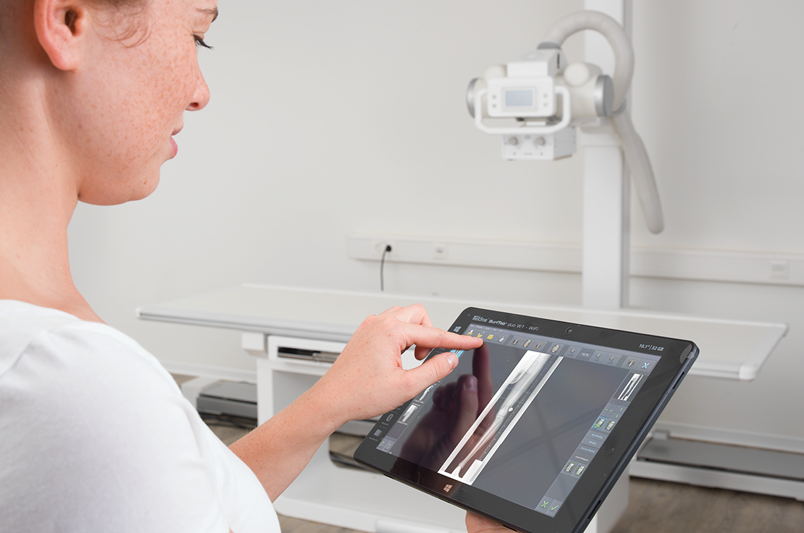
Stitching is traditionally used in radiology to produce whole-leg and whole-spine images. Only with this technology it is possible to visualise the entire apparatus with cassette or detector systems. It is often stated that X-ray stitching requires manual or semi-automatic overlapping and adjustment processes. However, if the user utilises a modern software solution, it takes over this task and automatically arranges the individual images using markers and a lead ruler. In addition, the overlapping areas in the individual images are visualised before stitching.
In CT, stitching is important for the visualisation of vascular trees or tumour expansions. By stitching several CT images, a coherent image of the entire vascular tree is created, which enables a precise diagnosis of stenoses or aneurysms. For example, the arterial supply of an organ or tissue can be reliably assessed. Another field of application is MRI imaging of the brain to examine tumours. By stitching several MRI slices, the radiologist obtains a more comprehensive representation of the tumour volume. This in turn is crucial for surgical planning or monitoring the progress of tumours.
The advantages of stitching are obvious. Images processed in this way offer the radiologist a better spatial resolution, a more comprehensive representation of anatomical structures, the possibility of reducing artefacts and a more precise localisation of pathologies. All of this contributes to diagnostics and therapy and ultimately helps the patient.
Stitching technology will continue to improve with the further development of imaging and software algorithms. Increasingly automated stitching processes, for example, will speed up the image stitching process and increase accuracy. A further decisive step would be if stitching could be carried out in real time during image acquisition. This could further increase efficiency and clinical benefit. It is foreseeable that stitching will continue to play an important role in radiology in the future thanks to continuous innovation and technological advances.
