Detection of malignant lesions in women with high breast tissue density - pilot study on Breast-CT from AB-CT
By combining continuously rotating X-ray tubes with innovative single-photon counting detectors, spiral computed tomography (CT) scanners for breast cancer screening enable superimposition-free 3D imaging with high image resolution and short scan times. A current pilot study also shows that women with high breast tissue density in particular benefit from the use of the spiral CT nu:view from the company AB-CT. 1
Women with rather dense breast tissue have an increased risk of breast cancer. In addition, very dense breast tissue can ‘mask’ suspicious lesions and significantly impair the sensitivity of conventional mammography.2 The detection of malignant lesions based purely on morphological characteristics (shape, margin) is a challenge in women with very dense breast tissue.
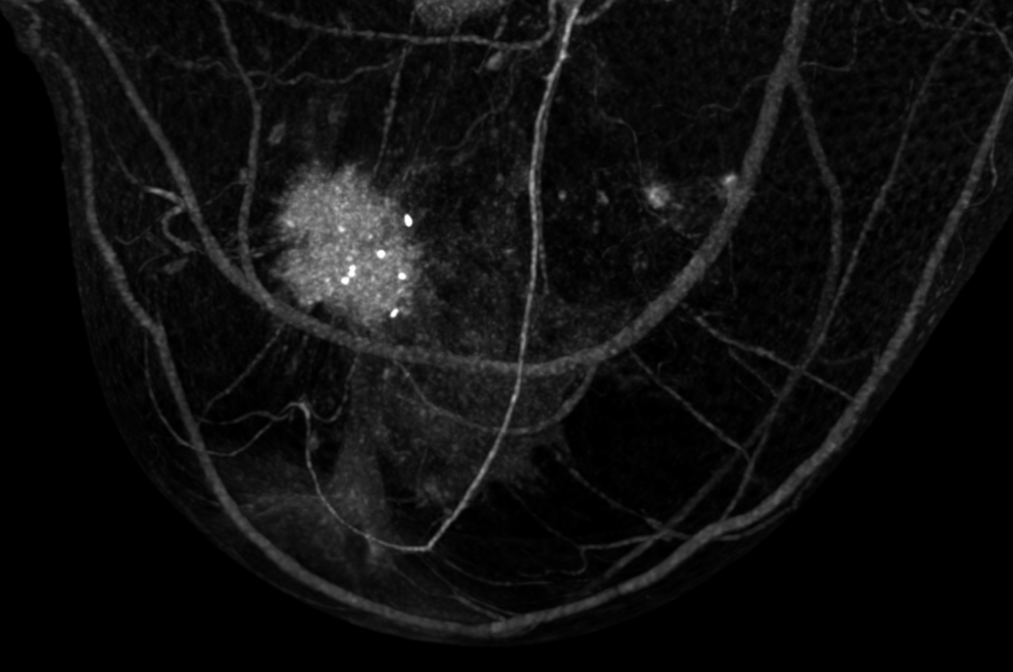
The advantages of spiral CT have so far mainly been seen in the superimposition-free 3D imaging and the improved patient comfort (examination without breast compression). Additionally, spiral CT can also precisely quantify lesion and tissue densities. The aim of the study by Weber et al.1 was therefore to differentiate malignant lesions from benign breast tissue, cysts and healthy but very dense breast tissue on the basis of tissue density.
The study included 40 women with the following previous findings:
- Malignant lesions: 12 women (median age 56.5 years)
- Fibroadenomas: 7 women (median age 51.0 years)
- Cysts: 12 women (median age 51.0 years)
- Extremely dense glandular tissue without findings: 9 women (median age 51.0 years)
The examination protocol included one breast CT scan with the nu:view device per breast without contrast injection. The image analysis to determine the median breast tissue density in Hounsfield units (HE; with interquartile range [IQR]) was performed independently by two examiners.
Significant differences between malignant lesions and other tissues
The two investigators determined density values of 60.2 (53.3-67.3) HE and 62.5 (55.67-76.3) HE for malignant lesions. For fibroadenomas, the density was 46.3 (41.9-59.5) HE and 44.5 (40.5-59.8) HE, for cysts 35.3 (24.3-46.0) HE and 39.7 (26.7-52.0) HE and for very dense breast tissue without clinical findings 28.7 (24.2-33.0) HE and 33.3 (31.7-36.8) HE.
These tissue densities differed significantly between malignant lesions and dense breast tissue without findings (p < 0.001), between malignant lesions and cysts (p < 0.001) and between fibroadenomas and dense breast tissue (p ≤ 0.003; see fig. 1).

Differentiation between different lesions and dense breast tissue without clinical findings with significant differences in tissue density.
High quality of findings and reproducibility
To assess the test quality, the area under the curve was determined for the differentiation between malignant lesions and other tissues (AUC with 95% confidence interval [CI]). The AUC values of both investigators showed a high quality of findings (AUC 1: 0.925 [CI 0.858-0.993]; AUC 2: 0.942 [CI 0.884-1.00]).
For the detection of malignant lesions, this resulted in a sensitivity of 91.7%-100% (depending on the cut-off) with a specificity of up to 81.4%. For the differentiation of malignant lesions from fibroadenomas, sensitivity and specificity were 75.0% and 70.0% respectively. The intraclass correlation, which quantified the diagnostic agreement between the two examiners (inter-reader), showed an almost perfect reproducibility of 0.978 (CI 0.961-0.988).
Conclusion
In summary, malignant lesions show significantly higher density values in examinations with the nu:view breast CT than the other examined tissues (except fibroadenomas). This means that breast CT can reliably detect malignant lesions even with high breast tissue density and differentiate them from cysts and dense breast tissue without clinical findings.
The ability to reliably detect this difference even without a contrast agent and with a short scan time could represent a significant advantage for women with high breast density compared to other methods such as contrast-enhanced mammography.
Sources
1Weber J, et al. Potential of non-contrast spiral breast CT to exploit lesion density and favor breast cancer detection: A pilot study. Eur J Radiol 2024,178, 111614.
2Freer PE. Mammographic breast density: impact on breast cancer risk and implications for screening. Radiographics 2015,35, 302-15.